sexta-feira, 21 de dezembro de 2012
sexta-feira, 30 de novembro de 2012
Resposta!
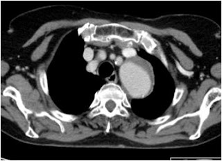
Atelectasis is defined as the collapse or closure of the lung resulting in reduced or absent gas exchange. It may affect part or all of one lung. It is a condition where the alveoli are deflated, as distinct from pulmonary consolidation.
terça-feira, 27 de novembro de 2012
sexta-feira, 23 de novembro de 2012
segunda-feira, 19 de novembro de 2012
segunda-feira, 12 de novembro de 2012
Caso
Doente de 70 anos, com neoplasia hematológica, submetido a esplenectomia.
Reavaliação abdominal por ecografia 24h após a cirurgia.
quinta-feira, 8 de novembro de 2012
segunda-feira, 22 de outubro de 2012
Resposta ao Caso Clínico
Adenoma da SUPRA-RENAL
- Incidence in the population is 2-8%
- Diagnosis is often made as an incidental finding on CT examination.
- In patient with no known primary, an adrenal mass is almost always a benign adenoma
- In a patient with a known neoplasm, especially lung cancer, an adrenal mass is problematic and diagnosing a metastasis versus an adenoma is critical for prognosis
Imaging findings
- CT
- Size greater than 4 cm tend to be metastases or adrenal carcinoma
- Heterogeneous appearance and irregular shape are malignant characteristics
- Homogeneous and smooth are benign characteristics.
- Intracellular lipid in adenoma results in low attenuation on CT
- Little intracytoplasmic fat in metastases results in high attenuation on non-enhanced CT
- Non-enhanced CT (NECT)
- Threshold 10 HU
- Sensitivity 79%, specificity 96%
- Contrast-enhanced CT (CECT)
- Because majority of CT examinations in oncology use IV contrast, the % washout is useful after 10 minutes.
- Adenomas have greater than 50% washout after 10 minutes
- Washout can also be used on adrenal masses that measure > 10 HU on NECT
- Alternative is to do MR or PET
- Size greater than 4 cm tend to be metastases or adrenal carcinoma
- MR
- Chemical Shift
- Most sensitive method for differentiating adenomas from metastases
- Sensitivity 81-100%. Specificity 94-100%.
- The difference in resonance rate of protons in fat and water is exploited in chemical shift.
- Intracellular lipid and water in same voxel result in summation of signal on "in-phase" and canceling out of signal on "out of phase"
- Spleen or muscle is used as an internal standard to visually quantify signal drop-off
- Liver is not a reliable standard because of steatosis
- Chemical Shift
segunda-feira, 15 de outubro de 2012
Caso Clínico
segunda-feira, 1 de outubro de 2012
quinta-feira, 27 de setembro de 2012
quarta-feira, 19 de setembro de 2012
segunda-feira, 17 de setembro de 2012
sexta-feira, 14 de setembro de 2012
segunda-feira, 10 de setembro de 2012
segunda-feira, 3 de setembro de 2012
terça-feira, 31 de julho de 2012
sexta-feira, 6 de julho de 2012
Practical MMR Mammography (Hardback)
Just Released! April 2012
Acclaim for the first edition: A handy reference of MRI findings for practicing radiologists in their daily work. Indications for breast MRI are excellently presented. Strongly recommended. Acta Radiologica Interesting and instructive book [...] the author successfully presents, evaluates and discusses the use of MR in the imaging of the breast [...]. Each chapter is enriched by numerous, clear and demonstrative illustrations [...] should be in the hands of all radiologists who practice mammography [...] and those who should know when and why MR mammography should be performed.
terça-feira, 3 de julho de 2012
sábado, 30 de junho de 2012
Resposta ao caso clinico
Duodenal diverticula are out-pouchings from the duodenal wall (intra luminal diverticulum discussed separately). They may result from mucosal prolapse or the prolapse of the entire duodenal wall and can be found at any point in the duodenum.
Demographics and clinical presentation
Duodenal diverticula are very common, found in up to 23% of asymptomatic patients, and in the vast majority remain asymptomatic throughout life. In 10% of patients, some symptoms are attributable to them, with only a minority requiring surgical intervention 2. Potential complications include : duodenal diverticulitis haemorrhage perforation abscess formation Diverticula located at the ampulla of Vater may cause difficulty for endoscopists as they attempt to cannulate the biliary system.
Pathology and classification
There are a two of types of duodenal diverticula: primary diverticulum secondary diverticulum A primary duodenal diverticulum occurs where there is prolapse of mucosa through the muscularis propria. They usually occur within the 2nd part (62%) and less commonly in the 3rd (30%) and 4th (8%) parts. Unlike secondary diverticula they are rarely seen in the 1st part. When they occur in the 2nd part, most (88%) are seen on the medial wall around the ampulla, 8% are seen posteriorly and 4% on the lateral wall. A secondary duodenal diverticulum results from prolapse of the entire duodenal wall and almost invariably occurs in the 1st part of the duodenum. These are true diverticula and are usually secondary to duodenal or peri duodenal inflammation.
quarta-feira, 27 de junho de 2012
quarta-feira, 13 de junho de 2012
domingo, 10 de junho de 2012
Avoiding Errors in Radiology
In Avoiding Errors in Radiology: Case-Based Analysis of Causes and Preventive Strategies, the authors provide 118 real-life examples of interpretation errors and wrong decisions from both diagnostic and interventional radiology. In each case, the authors discuss in detail the context in which the errors were made, the resulting complications, and strategies for future prevention. The cases are organized by body region, beginning with the cranium and then moving to cases of the breast, chest and abdomen, spinal column, musculoskeletal and vascular systems.
Features: *118 case studies facilitate analysis and discussion of causes of errors and offer preventive strategies to transfer into daily practice *956 high-quality images and explanatory drawings illustrate the cases and pinpoint errors of interpretation and in decision making Avoiding Errors in Radiology is a must-have reference for anyone involved ininterpreting images for diagnosis and in making decisions in interventional radiology.
quinta-feira, 31 de maio de 2012
Resposta ao Caso de Urgência - Incidentaloma
A ureterocele is a congenital abnormality found in the urinary bladder. In this condition called ureteroceles, the distal ureter balloons at its opening into the bladder, forming a sac-like pouch. It is most often associated with a duplicated collection system, where two ureters drain their respective kidney instead of one. Simple ureteroceles, where the condition involves only a single ureter, represents only twenty percent of cases. Ureteroceles affects one in 4,000 individuals, at least four fifths of whom are female. Patients are frequently Caucasian.
Since the advent of the ultrasound, most ureteroceles are diagnosed prenatally. The pediatric and adult conditions are often found only through diagnostic imaging performed for reasons other than suspicious ureteroceles.
quarta-feira, 23 de maio de 2012
terça-feira, 15 de maio de 2012
Delegação RADINT_IPOL@ CNR 2012
Posters
T1154 - Pleura e Revestimento Interno da Parede Torácica em Imagem
Abreu, Elisa de Melo; Palmeiro, Marta Morna; Marques, Vasco; Vasconcelos, Maria Antónia; Vinhais, Sofia
T1096 - Infeção Fúngica Pulmonar em Doentes Hemato-Oncológicos: Apresentações Imagiológicas
Morna Palmeiro, Marta; Melo Abreu, Elisa; Ip, Joana; Conceição e Silva, João Paulo
T1057 - Lesões Malignas do Mediastino: Revisão Pictórica
Morna Palmeiro, Marta; Melo Abreu, Elisa; Vasconcelos, Maria Antónia; Conceição e Silva, João Paulo
T1094 - Tumores Ósseos: Características em Radiologia Convencional
Morna Palmeiro, Marta; Melo Abreu, Elisa; Loureiro, Ana; Conceição e Silva, João Paulo
T1211 - As várias faces do Osteossarcoma: revisão pictórica
Vasconcelos, Maria Antónia; Abreu, Elisa; Palmeiro, Marta; Niza, João; Ip, Joana; Marques, Vasco
T1009 - Avaliação imagiológica após crioterapia renal - revisão de 21 casos
Joana Ip, Rui Carneiro, Isabel Duarte
T1010 - Estadiamento T1 e T2 do Cancro do Pulmão Não Pequenas Células - Avaliação da dimensão da TC versus resultado da peça operatória
Joana Ip, Catarina Callé, Susana Esteves, Nuno Abecasis, Fernando Cunha, Maria Teresa Almodovar, Isabel Duarte
T1011 - Cancro do Pulmão Não Pequenas Células: Associação entre características morfológicas de TC e o tipo histológico - revisão de 77 casos
Joana Ip, Catarina Callé, Susana Esteves, Nuno Abecasis, Fernando Cunha, Maria Teresa Almodovar, Isabel Duarte
Comunicações Livres
T1084 - Imagiologia dos Hilos Pulmonares
Abreu, Elisa de Melo; Palmeiro, Marta Morna; Vasconcelos, Maria Antónia; Vinhais, Sofia
T1054 - Lipossarcomas Musculoesqueléticos: Espectro Imagiológico e Correlação Anátomo-Patológica
Morna Palmeiro, Marta; Melo Abreu, Elisa; Conceição e Silva, João Paulo; Rito, Miguel
T1078 - Sarcoma de Ewing: Análise Clínico/Radiológica de uma Série
Abreu, Elisa de Melo; Palmeiro, Marta Morna; Vasconcelos, Maria Antónia; Vinhais, Sofia
T1225 - Quistos simples do ovário - que fazer?
Vasco Marques, Teresa Margarida Cunha
quarta-feira, 9 de maio de 2012
terça-feira, 1 de maio de 2012
terça-feira, 24 de abril de 2012
segunda-feira, 9 de abril de 2012
CIRSE | Cardiovascular and Interventional Radiological Society of Europe
sábado, 7 de abril de 2012
quinta-feira, 5 de abril de 2012
terça-feira, 3 de abril de 2012
segunda-feira, 2 de abril de 2012
sexta-feira, 30 de março de 2012
segunda-feira, 26 de março de 2012
domingo, 25 de março de 2012
quarta-feira, 21 de março de 2012
sábado, 17 de março de 2012
terça-feira, 13 de março de 2012
Subscrever:
Comentários (Atom)


.jpg)


